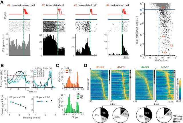
Figure 3.
Different types of functional activity in the M1 and M2 neurons. A, Definition of task-related activity. The number of spikes during contralateral and ipsilateral movement trials was plotted against task relevance index for individual neurons (right scatter plot; see Materials and Methods). Black and gray dots indicate the task-related (blue line, p < 10−6; blue dashed line, ≥250 spikes) and non–task-related (discarded) neurons, respectively. Red numbers correspond to examples of activities (left panels). Top, Middle, and Bottom, Pedal trajectories, spike raster plots, and PETHs of preferred movement, respectively. Bin width, 20 ms. B, Categorization of Hold- and Pre-go-type activities by dependence on holding time. PETHs calculated from the different holding time trials (top). Intersection with criterion (red dashed line, 75% of activity in an averaged PETH) was plotted on the holding time, and the slope value of the regression was obtained (bottom). If a neuron had Hold-type-related activity, the slope value was negative (left column). By contrast, the slope value was ∼0 if neuronal activity was independent of holding time (right column). C, Distribution of slope in the motor cortices. Histograms of slopes exhibit a clear bimodality in both M1 and M2. D, Three types of task-related activity in RS and FS neurons in the motor cortices. Top, Each row shows normalized Gaussian-filtered PETH (σ = 12.5 ms, in 0.05 ms bins) for a single neuron (aligned with the onset of choice: vertical line at 0 s). The task-related neurons were sorted by the order of peak time (early to late). Hold-, Pre-go-, and Post-go-type activities are indicated on the right side. Bottom, Population ratios of different activity types for RS and FS neurons in the motor cortices. Black, gray, and white represent Hold-, Pre-go-, and Post-go-type activity, respectively. ***p < 0.001 (2 × 2 χ2 tests).

