Figure 6.
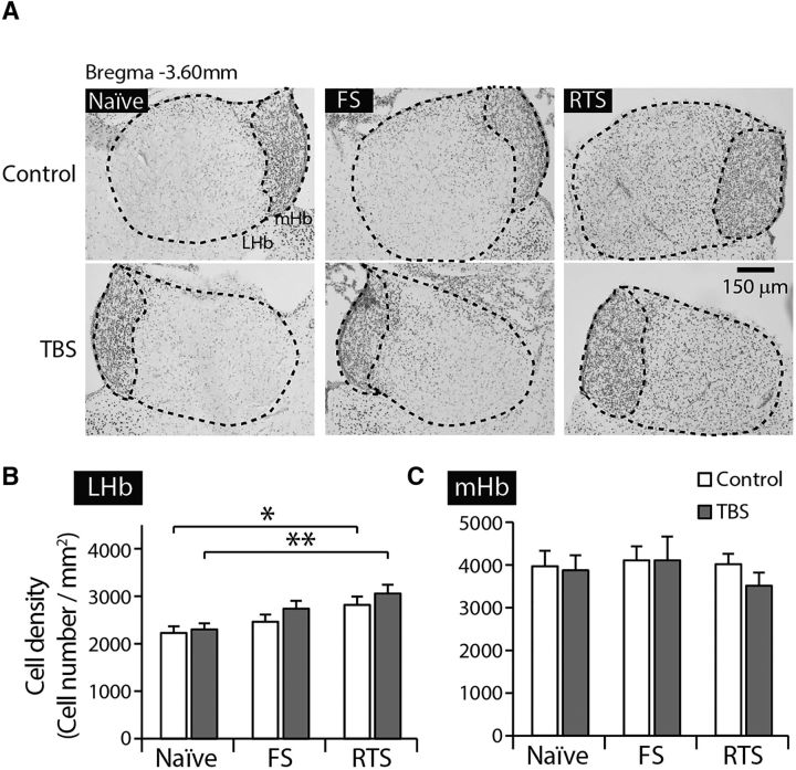
Stress exposure increased the density of neurons expressing phosphorylated CREB in the LHb. A, Representative images showing pCREB immunoreactivity in the habenular complex of naive, FS-, and RTS-exposed animals. B, The density of pCREB-expressing cells in the LHb was significantly increased upon exposure to stressors (two-way ANOVA, F(2,114) = 9.37, p < 0.001). Stress exposure per se increased pCREB-positive cell density in the LHb in the absence of any stimulation (F(2,57) = 3.85, p < 0.05, one-way ANOVA with Bonferroni post hoc analysis). C, No significant differences in the density of pCREB-positive cells were observed in the mHb upon stress exposure (two-way ANOVA, F(2,114) = 0.42, p > 0.7) or TBS (two-way ANOVA, F(1,114) = 0.45, p > 0.5). *p < 0.05. **p < 0.01.

