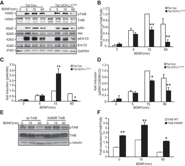
Figure 9.
Tat-UCH-L175–85 causes decreased activation of TrkB and its downstream signaling pathways. A, Representative images of immunoblots showing BDNF-induced phosphorylation of TrkB, Akt, and ERK1/2 in cultured hippocampal neurons under the indicated conditions. Neurons were pretreated with Tat-Con (10 μm, 30 min) or Tat-UCH-L175–85(10 μm, 30 min) before BDNF (50 ng/ml) stimulation. B–D, Quantitative analysis of TrkB (B), Akt (C), and ERK1/2 (D) activation in neurons under Tat-UCH-L175–85 and Tat-Con incubation. p-TrkB, p-ERK1/2, and p-Akt levels were normalized to the phosphorylation levels of BDNF alone treatment group detected at the 15 min time point. Graphs represent means ± SEM. n = 4, *p < 0.05, **p < 0.01, one-way ANOVA. E, Representative images of immunoblots showing BDNF-induced phosphorylation of TrkB in HEK293 cells transfected with WT or K460R TrkB for the indicated times. F, Quantitative analysis of TrkB activation. Graphs represent means ± SEM. n = 3, *p < 0.05, **p < 0.01, one-way ANOVA.

