Figure 2.

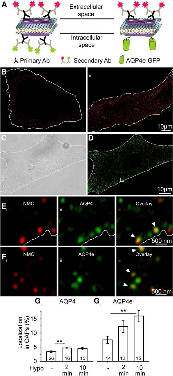
AQP4e is localized in OAPs, and it promotes their formation in hypoosmotic conditions. A, Schematically depicted double labeling of AQP4 channels at the plasma membrane OAPs. Primary NMO antibodies label the extracellular site of AQP4s (secondary antibodies are conjugated to a red fluorescent dye) and are thus markers of OAPs, whereas primary commercial antibodies label OAPs at the intracellular domain of the AQP4 channel (secondary antibodies are conjugated to a green fluorescent dye). Recombinant AQP4e isoforms are tagged with GFP. Of note, commercial pan antibodies also label intracellular MiDs containing AQP4s (see Eiii). B, Fluorescent micrographs of astrocytes labeled with NMO primary antibodies and delineated with white lines. Bi shows an equatorial plane of NMO-labeled impermeabilized astrocytes, where NMO labeling is restricted to the cell surface. We used this type of labeling to assess OAPs. Bii shows an equatorial plane of NMO-labeled formaldehyde-permeabilized astrocytes, where NMO labeling is observed also in the cytoplasm. C, DIC image of a cultured rat astrocyte. The cell is delineated with the white line. D, SIM fluorescence micrograph of the same astrocyte as in B, labeled as schematically shown in A. The squared area is enlarged in E, displaying OAPs extracellularly labeled with NMO antibodies (Ei), intracellularly labeled with commercial AQP4 antibodies (Eii), and the overlay of both signals (Eiii). The white line represents the edge of the astrocyte, as determined on the DIC image. F, Section of an astrocyte showing OAPs extracellularly labeled with NMO antibodies (Fi), the intracellular signal of AQP4e-GFP (Fii), and the overlay of both signals (Fiii). The arrowheads point to colocalized fluorescent signals of OAPs, labeled by NMO antibodies, that were either colabeled by AQP4 antibodies (Eiii) or AQP4e-GFP (Fiii). G, OAPs represented 3.4 ± 0.3% of all antibody-labeled AQP4 microdomains in untransfected cells. Stimulation with Hypo solution increased the percentage of OAPs to 4.7 ± 0.3% (2 min) and to 4.5 ± 0.5% (10 min), respectively (Gi). Of all recombinant AQP4e-GFP MiDs in the transfected cells, 7.6 ± 1.3% were detected in OAPs. Gii, Stimulation with Hypo increased the percentages to 12.3 ± 2.2% (2 min) and to 15.9 ± 2.0% (10 min). Numbers in the bars denote the number of cells analyzed. **p < 0.01.
