Figure 3.
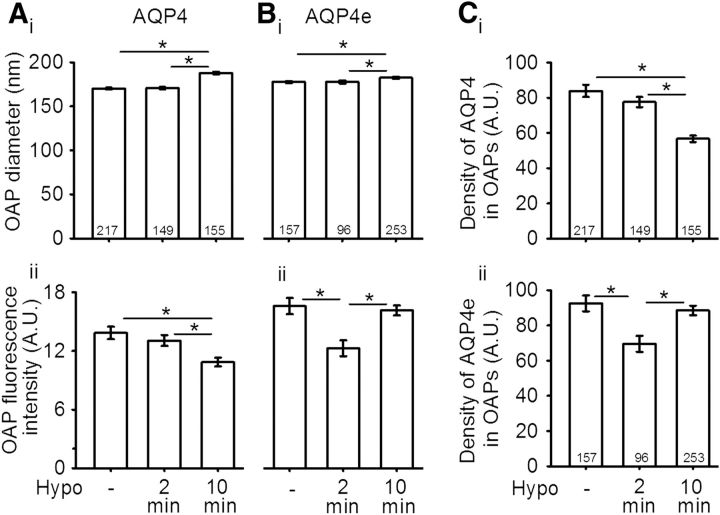
Hypotonicity increases the average OAP diameter in astrocytes. Ai, The average diameter of OAPs (in nanometers) in rat astrocytes intracellularly colabeled with AQP4 antibodies was 170 ± 1 in isoosmotic conditions. After 2 min of Hypo stimulation, it remained virtually unchanged (171 ± 1) and increased to 188 ± 1 after 10 min of Hypo stimulation. The corresponding average fluorescence intensity in arbitrary units (A.U.) is shown in Aii; it decreased from 13.8 ± 0.6 in isoosmotic conditions to 13.1 ± 0.6 and 10.9 ± 0.5 after 2 min and after 10 min of Hypo, respectively. Bi, The average diameter of OAPs (in nm) containing GFP-tagged AQP4e isoform was 178 ± 1 in isoosmotic conditions. After 2 min of Hypo stimulation, the average diameter did not change (178 ± 2), while it increased to 183 ± 1 after 10 min of Hypo stimulation. Bii, The average fluorescence intensity (in A.U.) of the same OAPs as in Bi was 16.6 ± 0.8 in isoosmotic conditions, transiently decreased to 12.3 ± 0.8 after 2 min of Hypo stimulation, and then increased to 16.1 ± 0.5 after 10 min. C, The AQP4 density was measured as the ratio between the average fluorescence intensity of OAP and the OAP diameter. The density of AQP4 (in A.U.) in OAPs (Ci) was 83.8 ± 3.5 in controls and decreased to 77.5 ± 2.9 (after 2 min Hypo stimulation) and to 56.6 ± 1.9 (after 10 min of Hypo stimulation). Cii, The decrease in the density of AQP4e in OAPs was transient, from 92.5 ± 4.5 in controls, to 69.5 ± 4.6 (after 2 min of Hypo stimulation), and to 88.5 ± 2.7 (after 10 min of Hypo stimulation). Numbers in the bars or brackets represent the number of OAPs analyzed. *p < 0.05.

