Figure 3.
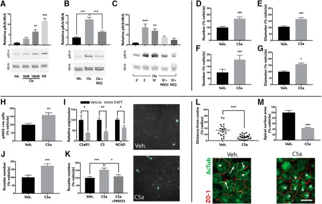
C5aR1 signals through PKCζ to maintain cell polarity in vitro. A, Mouse neurosphere cultures demonstrate C5a concentration-dependent p42/44 phosphorylation. B, The response to 100 nm C5a is prevented by PKCζ inhibition. C, Human rosette cultures demonstrate time-dependent p42/44 phosphorylation to 10 nm hC5a. The response is prevented through pretreatment with C5aR1-A or PKCζ inhibition. D–G, Mouse and human neurosphere cultures dissociated and grown over a 7 d period demonstrate an increase in number (D, F) and diameter (E, G) in response to C5a. H–K, Treatment of human rosettes with 10 nm C5a. H, C5a increases M-phase-positive cells in neural rosettes as determined by pHH3 immunocytochemical analysis. I, DAPT (gray bars) treatment induced loss of rosettes and a decrease in mRNA of NCAD, C5AR1, and C5 compared with vehicle (black bars) treatment. J, K, Maintenance of rosette architecture after single-cell dissociation (K) or DAPT treatment (J) was promoted by exogenous C5a addition. Adjacent images are representative of DAPT-treated rosettes in the presence or absence of C5a. NCAD (white) and computational outlines (green) of rosette apical lumens are shown. L, M, NE-4C cells grown on transwell membranes demonstrate reorganization of the mitotic spindle (L), as determined by acetylated tubulin staining (green), and reduction in apical surface area (M), outlined by ZO-1 (red) in response to C5a. White arrows are representative distances from mitotic spindle to calculated cell center as shown in L. Scale bar, 20 μm. Veh., Vehicle. *p ≤ 0.05, **p ≤ 0.01, ***p ≤ 0.001.

