
Figure 4.
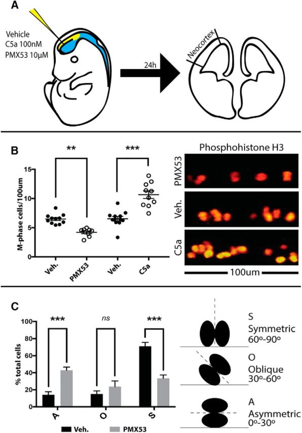
C5aR1 signaling alters neural progenitor division planes and proliferation in vivo. A, Schema of the in utero injection process. Briefly, 1 μl of 100 nm mC5a, 1 μm PMX53, or vehicle was delivered to the embryonic ventricle in utero. After 24 h, brains were processed for immunohistochemistry. M-phase cells, as determined by pHH3 staining, were counted along the ventricular surface of the neocortex. B, In utero injection of mC5a to the embryonic ventricle increases, whereas blockade of C5aR1 signaling using PMX53 decreases, the number of M-phase apical progenitor cells. C, Cleavage plan analysis demonstrates a shift from symmetric division toward asymmetric division after treatment with C5aR1 antagonist. S, Symmetric division; A, asymmetric division; O, oblique division; Veh., vehicle. **p ≤ 0.01, ***p ≤ 0.001.