
Figure 1.
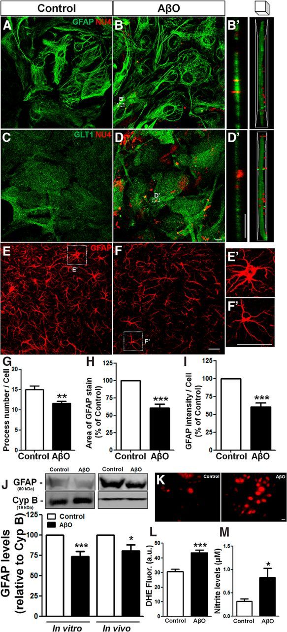
AβOs bind to astrocyte membranes, are internalized, and trigger astrocyte activation. Mouse hippocampal astrocyte cultures were exposed to vehicle or 500 nm AβOs for 24 h. Cells were then analyzed by immunocytochemistry for GFAP, GLT1, and AβOs (NU4 antibody immunoreactivity; A–D); representative z-stack orthogonal cut images of GFAP/NU4 (B′) and GLT1/NU4 (D′); levels of ROS (K, L) and nitrite production (M). E–I, AβO or vehicle was infused intracerebroventricularly in mice and GFAP immunostaining was analyzed in the hippocampus (E, F). Astrocyte morphology was evaluated by analyzing the number of processes (G), cellular area (H), and intensity of GFAP immunoreactivity per cell (I). GFAP levels were analyzed by Western blotting of cultured astrocytes or hippocampal tissue homogenates (J). Exposure to AβOs triggered astrocyte activation in vitro and induced astrocytic atrophy in vivo. Scale bars: D, D′, K, 10 μm; F, F′, 30 μm. *p < 0.050, **p < 0.010, and ***p < 0.001; n = 3–6 experiments with independent astrocyte cultures and three animals per experimental group; 30–45 cells were analyzed per experimental condition. Student's t test.