
Figure 4.
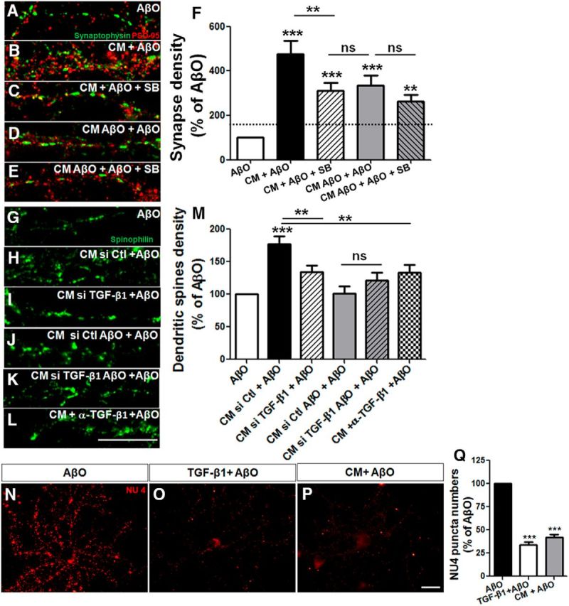
Astrocyte protection against AβOs is mediated by TGF-β1. Mature hippocampal neurons (14 DIV) were maintained for 30 min in the absence or presence of CM derived from astrocyte cultures (CM) or CM derived from astrocyte cultures previously primed by AβO (CM AβO), and were then exposed for 3 h to 500 nm AβOs. When present, 10 μm SB-431542 was added 30 min before CM addition. Synapse density was evaluated by double immunocytochemistry for PSD-95 and synaptophysin and determination of juxtaposed puncta (A–F). A–E, Representative images of dendritic segments under different experimental conditions. Density of dendritic spines were analyzed by spinophilin labeling in neurons cultured for 12 DIV and maintained in different CM plus AβOs. Neurons were treated with CM siRNA control (CM si Ctl), CM siRNA for TGF-β1 (CM siTGF-β1), or in the presence of neutralizing antibody against TGF-β1 (CM + α-TGF-β1). Representative dendrites of spinophilin labeling (G–L) and quantification of the density of dendritic spines (M). Prior addition of 10 ng/ml purified TGF-β1 to AβO-exposed neurons mimics the effect of CM and impairs AβO binding to neurons (measured by the quantification of the number of NU4 puncta; N–Q). Scale bars: 10 μm. **p < 0.010 and ***p < 0.001, one-way ANOVA followed by Tukey's post hoc tests. F, M, Q, n = 3–4 experiments with independent neuronal cultures. A total of 90–100 cells (F), 37–40 cells (M), and 60–95 cells were analyzed per experimental condition (Q).