
Figure 3.
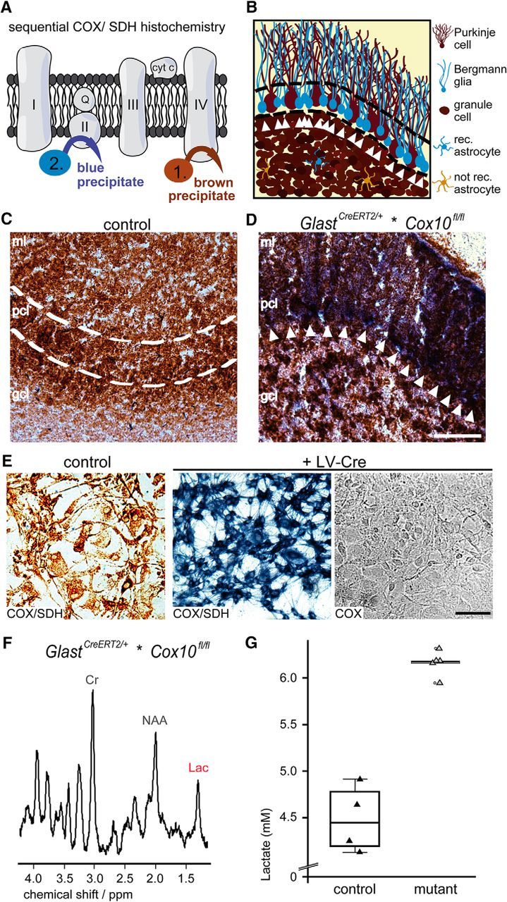
Sequential COX/SDH histochemistry proving the loss of COX activity in BG cells. A, Identification of respiration deficient (COX neg.) cells by sequential COX/SDH histochemistry. In control cells, COX activity generates a brown precipitate and prevents the emergence of a blue precipitate by the SDH reaction, which therefore marks mutant cells. B, Scheme illustrating mutant BG in the PC layer (blue, arrowheads) next to PCs (brown, COX active) as shown below. C, In control mice, mitochondrial COX activity leads to a brown precipitate in all cells of the cerebellum (age 14 months). D, In age-matched mutant mice, the blue precipitate marks BG cells (white arrows). Note also blue spots in the granule cell layer indicating astrocytes. Scale bar, 50 μm, E, Lentiviral-mediated deletion of Cox10 in cultured astrocytes maintained for 8 weeks in vitro leads to inactive cytochrome c oxidase in all astrocytes, as detectable by the emergence of a blue precipitate and the absence of a brown precipitate when only COX medium was applied. Scale bar, 100 μm. F, Localized proton MR spectra obtained from the cerebellum (voxel size: 4 × 1 × 2 mm3) revealed increased lactate concentrations in GlastCreERT2/+*COX10fl/fl mice. G, When quantified, mutant mice revealed a concentration of 6.2 ± 0.2 mm (controls 4.5 ± 0.4 mm). Lac, lactate; Cr, creatine; NAA, N-acetylaspartate; rec., Cre recombined; ml, molecular layer; pcl, PC layer; gcl, granule cell layer.