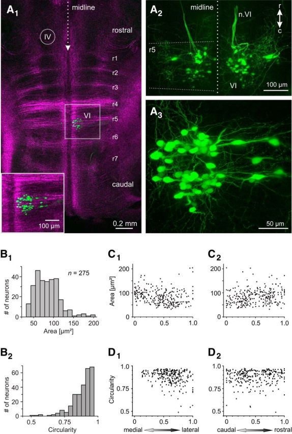
Figure 1.

Anatomical organization of the abducens nucleus in larval X. laevis. A, Horizontal confocal reconstruction of a hindbrain whole-mount preparation of a stage 54 tadpole depicting retrogradely labeled abducens motoneurons in r5 after unilateral (A1, A3) or bilateral (A2) application of Alexa Fluor 488 dextran to the VIth motor nerve(s) at the level of the LR muscle; brainstem-crossing fibers, visualized by 633 nm illumination (magenta-colored crossing fibers in A1), indicate the center of r1–7, respectively; inset in A1 is a higher magnification of the outlined area (VI). The location of the trochlear nucleus (IV) is indicated in A1. Note that the two green fiber bundles in A2 represent the bilateral VIth motor nerves (n.VI) traversing rostrally beneath the ventral surface of the hindbrain. r, rostral; c, caudal. B, Somal size (cross-sectional area, B1) and circularity (B2) distributions of retrogradely labeled abducens motoneurons (n = 275); circularity of 1 indicates a round cell body. C, D, Dependency of somal size (C) and circularity (D) on the mediolateral (C1, D1) and rostrocaudal (C2, D2) motoneuronal position within the abducens nucleus; positions were normalized to the most medial/rostral (0) and most lateral/caudal (1) part of the nucleus, respectively.
