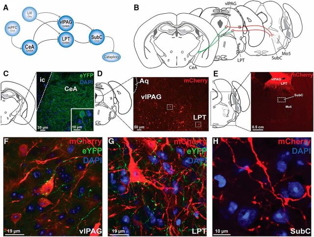
Figure 10.
Circuits controlling cataplexy. A, Positive emotions activate cortical areas such as the mPFC, which in turn activate the CeA. GABA cells in the CeA then inhibit midbrain regions, including the LPT and vlPAG, which normally function to inhibit the atonia-generating network in the brainstem (i.e., SubC). Inhibition of these midbrain regions disinhibits the muscle paralysis circuit during cataplexy. B, Mapping of the cataplexy circuit using virally tractable tracers. rAAV5/EF1a-DIO-ChETA-eYFP and rAAV8/hSyn-DIO-mCherry were injected into the CeA and vlPAG/LPT, respectively, of orexin−/−,VGAT-Cre mice. C, GABA CeA neurons expressed eYFP. D, GABA vlPAG/LPT neurons expressed mCherry. E, GABA vlPAG/LPT neurons expressing mCherry send projections to the SubC region. F, GABA CeA fibers expressing eYFP are located in the vicinity of a GABA vlPAG cell body expressing mCherry. G, GABA CeA fibers expressing eYFP are located in the vicinity of a GABA LPT cell body expressing mCherry. H, GABA vlPAG/LPT projections are located within the SubC region. Aq, Aqueduct; Mo5, trigeminal motor nucleus; ic, internal capsule; mPFC, medial prefrontal cortex; LH, lateral hypothalamus; Orx, orexin; Glut, glutamate.

