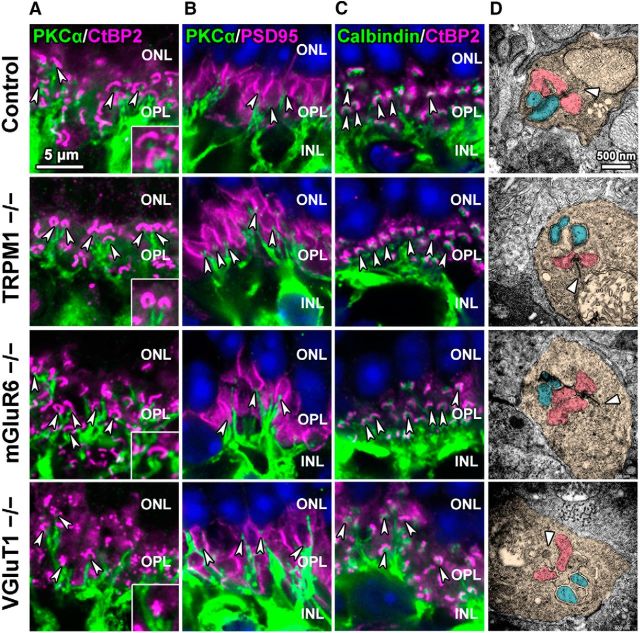
Figure 4.
Synaptic connections in the OPL are maintained in TRPM1−/−, mGluR6−/−, and VGluT1−/− retinas. A–C, Immunohistochemical analysis of the 1M control, TRPM1−/−, mGluR6−/−, and VGluT1−/− mouse retinas using retinal cell marker antibodies as follows: A: PKCα (green) and CtBP2 (magenta); B: PKCα (green) and PSD95 (photoreceptor terminal; magenta); C: calbindin (horizontal cell; green) and CtBP2 (magenta). Nuclei were stained with Hoechst (blue). Arrowheads in A and C indicate rod bipolar dendrites (A) or horizontal cell processes (C) in the vicinity of photoreceptor ribbons. Arrowheads in B indicate invaginations of rod bipolar cells into photoreceptor terminals (B). ONL, Outer nuclear layer. Insets represent OPL regions at high magnification. D, Ultrastructural analysis of photoreceptor ribbon synapses in the OPL of control, TRPM1−/−, mGluR6−/−, and VGluT1−/− mouse retinas at 1M by electron microscopy. Arrowheads indicate synaptic ribbons. Photoreceptors are tinted orange. Horizontal cell processes are tinted pink. Bipolar cell dendritic terminals are tinted blue.

