
Figure 5.
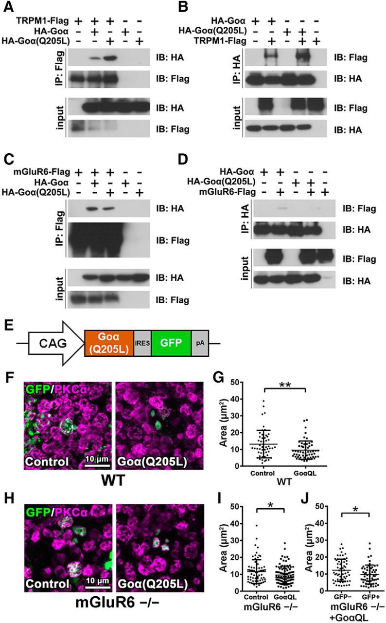
Effect of Goα overexpression on axon terminal formation of rod bipolar cells. A, B, Interaction of TRPM1 with the Goα subunit. The FLAG-tagged TRPM1 expression plasmid was transfected with HA-tagged Goα or HA-tagged Goα(Q205L) expression plasmids into HEK293T cells (A, B). The cell lysates were subjected to immunoprecipitation with either an anti-FLAG (A) or an anti-HA antibody (B). Immunoprecipitated proteins were analyzed by Western blotting with anti-FLAG and anti-HA antibodies. C, D, Interaction of mGluR6 with the Goα subunit. The FLAG-tagged mGluR6 expression plasmid was transfected with HA-tagged Goα or HA-tagged Goα(Q205L) expression plasmids into HEK293T cells (C, D). The cell lysates were subjected to immunoprecipitation with either an anti-FLAG (C) or an anti-HA antibody (D). E, Schematic representation of the expression construct of Goα(Q205L) (pCIG-Goα(Q205L)) used for in vivo electroporation into P0 wild-type and mGluR6−/− mouse retinas. F, The pCIG (empty vector) or pCIG-Goα(Q205L) plasmids were electroporated into P0 wild-type mouse retinas. The retinas were harvested at 1M, spread flat, and immunostained with antibodies against GFP (green) and PKCα (magenta). The electroporated cells express EGFP mediated by the IRES sequence. G, Axon terminal areas of both EGFP- and PKCα-positive cells were measured. **p < 0.01 by unpaired Student's t test. Error bars represent ± SD from the means of n = 52 or 57 [control or Goα(Q205L), respectively] rod bipolar axon terminals collected from individual animals (n = 3). H, The pCIG (empty vector) or pCIG-Goα(Q205L) plasmids were electroporated into P0 mGluR6−/− mouse retinas. I, Axon terminal areas of both EGFP- and PKCα-positive cells were measured. *p < 0.05 by unpaired Student's t test. Error bars represent ± SD from the means of n = 67 or 96 [control or Goα(Q205L), respectively] rod bipolar axon terminals collected from individual animals (n = 3). J, Axon terminal areas of cells that were both EGFP- and PKCα-positive are compared with axon terminal areas of PKCα-positive cells without EGFP signals in Goα(Q205L) transfected retinas. *p < 0.05 by unpaired Student's t test. Error bars represent ± SD from the means of n = 60 or 59 (EGFP-negative or EGFP-positive, respectively) rod bipolar axon terminals collected from individual animals (n = 3). GoαQL, Goα(Q205L).