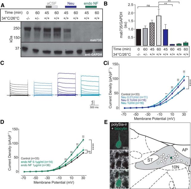
Figure 3.
polySia removal enhances current voltage relationship within NTS neurons. A, Representative Western blot using Mab735 to detect polySia during slice recovery, and following incubation in aCSF (gray/black box), Neu (0.1 U/ml, blue box), or endoNF (1 μg/ml, green box). GAPDH was used as a loading control. B, Comparison of the amount of protein detected using Mab735 during slice recovery (black and gray), and incubation in aCSF, Neu, or endoNF. polySia-ir was significantly reduced after 60 min incubation in Neu (blue box, p = 0.0039, n = 3) or 45 min incubation in endoNF (green box, p = 0.0042, n = 3). Data are normalized to GAPDH with the zero time point value set to 1. Enzymatic removal of polySia in NTS brain slices increases steady-state outward currents. C, Typical current traces (depolarizing voltage step: Δ10 mV) from NTS neurons incubated in aCSF (black), or Neu (0.01 U/ml, light blue, 0.1 U/ml, dark blue, and 1 U/ml, aqua, respectively). Ci, Grouped data reveal that Neu (0.1 and 1 U/ml) significantly increased current density of neurons at depolarized potentials ≥ 0 mV (p = 0.0002, n = 16 and n = 11, respectively). D, Grouped data show that incubation in endoNF at 1 μg/ml (dark green), but not 0.1 μg/ml (light green), significantly increased current density of neurons at depolarized potentials ≥ 0 mV (p < 0.0001, n = 36). Data are mean ± SEM. E, Recovered cell (green) within the NTS surrounded by polySia-ir puncta (white) coating the soma and proximal dendrite. The recovered cell was recorded while superfused with aCSF (control). Schematic coronal section showing distribution of recorded neurons in the NTS recovered following incubation in aCSF or endoNF. **p < 0.01; ****p < 0.0001; ns, p < 0.05.

