Figure 3.
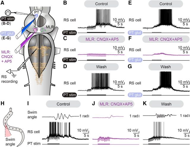
The descending glutamatergic drive from the PT to the MLR evokes reticulospinal activity and locomotion. A, In isolated brainstem preparations, glutamatergic antagonists were microinjected in the MLR. RS activity was recorded extracellularly in response to electrical (2 ms pulses, 5 Hz, 10 s train, 8–12 μA) or chemical stimulation (glutamate 2.5 mm, 20–80 ms pulses, 2 Hz, 9–11 pulses) of the PT. B–D, Microinjection of 36.4–78.4 pmol of CNQX (1 mm) and 18.2–39.2 pmol of AP5 in the MLR (0.5 mm) dramatically decreased RS activity elicited by PT stimulation (7 μA, 10 s train, 2 ms pulses, 4 Hz). Recovery was obtained after 62–108 min of washout. E–G, Microinjection of 51.5–119.9 pmol of CNQX (1 mm) and 25.7–156.4 pmol of AP5 (0.5 mm) in the MLR reduced RS activity elicited by PT chemical stimulation with 1.0–19.1 pmol of glutamate (illustrated case: 50 ms pulses, 2 Hz, 11 pulses). Responses recovered after 88–135 min of wash out. H, In a semi-intact preparation where RS neurons were recorded intracellularly to monitor the activity of brainstem locomotor circuits, glutamatergic antagonists were microinjected in the MLR. Angular variations (radians) of the curvature of a mid-body segment were measured over time during swimming. I–K, Microinjections of 34.8–68.4 pmol of CNQX (1 mm) and 17.4–34.2 pmol of AP5 (0.5 mm) in the MLR dramatically reduced RS activity and swimming movements elicited by PT stimulation (illustrated case: 10 μA, 10 s train, 2 ms pulses, 5 Hz). These effects were reversed after 48–135 min of washout. Data from B–D, E–G, and I–K are from three different preparations.

