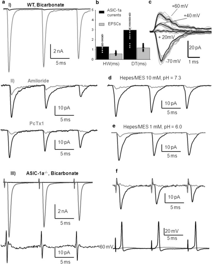
Figure 4.
Blockage of glutamatergic EPSCs reveals that protons released from presynaptic vesicles elicit postsynaptic ASIC-mediated currents and action potentials in MNTB neurons from WT mice. aI, Representative traces of EPSCs before (gray) and after (black) blocking AMPA, NMDA, GABA, and glycine receptors with CNQX (40 μm), d-APV (50 μm), bicuculline (20 μm), and strychnine (2 μm) in an MNTB neuron from a WT mouse [postnatal day 15 (P15)]. aII, Higher magnification of the ASIC-1a-mediated currents insensitive to CNQX, APV, bicuculline, and strychnine (black). Mean amplitudes were 46 ± 3 pA (n = 27). These ASIC-1a-mediated currents were highly reduced by amiloride (top, gray trace; n = 15) and were inhibited by PcTx1 (bottom, gray trace; n = 4). aIII, Top, Representative traces of EPSCs before (gray) and after (black) blocking AMPA, NMDA, GABA, and glycine receptors with CNQX (40 μm), d-APV (50 μm), bicuculline (20 μm), and strychnine (2 μm) in an MNTB neuron from ASIC1a−/− mouse (P16). Bottom, Higher-magnification image showing the absence of any current after postsynaptic receptor inhibition. b, Mean half-width and decay time of amiloride and PcTx1-sensitive ASIC-1a-mediated currents compared with glutamatergic EPSCs (HW: 1.31 ± 0.1 ms vs 0.59 ± 0.03 ms; DT: 3.0 ± 0.2 ms vs 1.18 ± 0.05 ms; n = 27; Student's t test, p < 0.001). c, Sample traces of ASIC-1a currents at different membrane potentials in WT mouse (P14). d, Increasing pH buffering using a 10 mm HEPES-based extracellular solution, pH 7.3, the ASIC-1a-mediated currents in MNTB neurons from WT mice were highly reduced (traces from a P15 mouse). e, ASIC-1a-mediated currents were highly diminished when the HEPES-based extracellular solution was maintained at pH 6.0 due to the desensitization of ASICs (traces from a P17 mouse). f, ASIC-1a-mediated currents (top, black trace) evoked by 100 Hz stimulation of presynaptic axons after the blockage of postsynaptic receptors with NBQX (a higher-affinity AMPA receptor antagonist, 10 μm) plus d-APV (50 μm), bicuculline (20 μm), and strychnine (2 μm) in a P15 WT mouse. Mean amplitudes were 38 ± 8 pA (HW, 1.1 ± 0.2 ms; DT, 2.7 ± 0.3 ms; n = 7). These currents were able to evoke APs (measured in current-clamp configuration at the resting potential of the MNTB neurons) whose amplitudes and kinetics did not differ from those evoked by glutamatergic currents (bottom, black trace; current-clamp recording). PcTx1 inhibited both ASIC-1a-mediated currents and evoked APs (gray traces).

