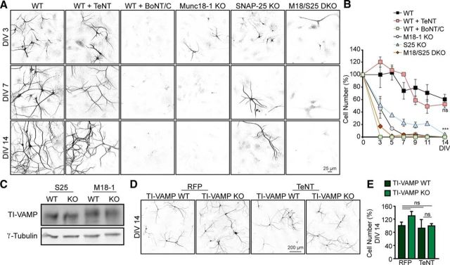
Figure 1.
Neuronal loss upon depletion of t-SNAREs and Munc18-1 but not v-SNAREs. A, Cortical neuronal cultures from WT, WT infected with TeNT or BoNT/C, Munc18-1 knock-out (M18–1 KO), SNAP-25 (S25) KO, and Munc18-1/SNAP-25 DKO (M18/S25 DKO) were fixed at different time points (DIV 3, 5, 7, 9, 11, and 14) and stained with a dendritic marker (MAP2). B, Quantification of number of neurons per 97 fields of view; not significant, p > 0.05. ***p < 0.001 (two-way ANOVA followed by the post hoc Bonferroni's test). C, E18 brain lysates from WT, M18–1 KO, and S25 KO were analyzed by Western blot and showed no differences in TI-VAMP levels between mutants and control animals (n = 3). D, Cortical neuronal cultures from TI-VAMP WT and TI-VAMP KO infected with TeNT or RFP (as control) were fixed at DIV 14 and stained with a dendritic marker (MAP2). E, Quantification of number of neurons per field of view. No differences in cell survival were found: not significant, p > 0.05 (one-way ANOVA followed by the post hoc Bonferroni's test). Data are mean ± SEM.

