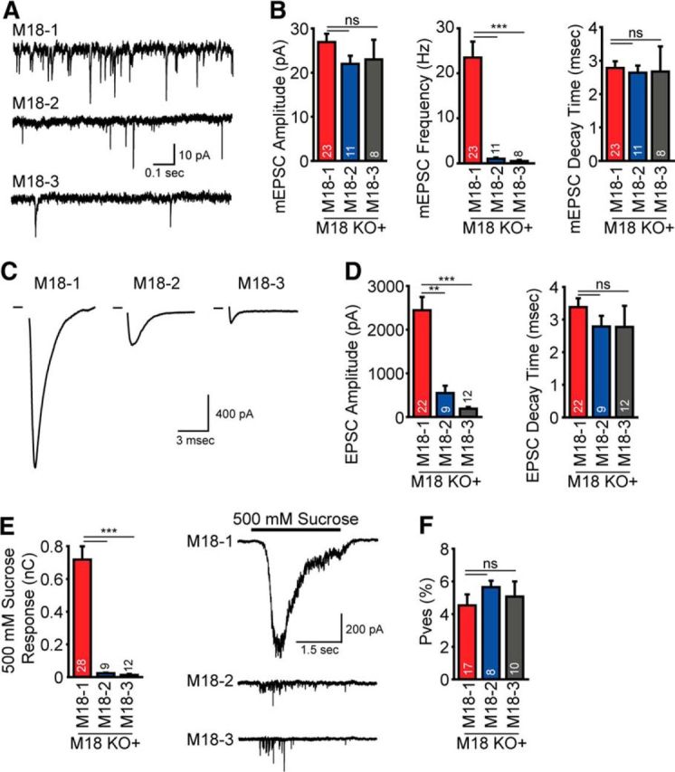
Figure 6.

Munc18-2/-3 expression does not rescue synaptic transmission in Munc18-1 KO neurons. A, Example traces of spontaneous release in neurons rescued with Munc18-1 (M18–1), Munc18-2 (M18–2), or Munc18-3 (M18–3). B, Spontaneous frequency is impaired in neurons expressing Munc18-2 or -3, whereas spontaneous amplitude and decay time are normal (amplitude: Munc18-1, 26.9 ± 1.9 pA; Munc18-2, 22.0 ± 1.8 pA; Munc18-3, 23.0 ± 4.5 pA; p > 0.05, frequency: Munc18-1, 23.5 ± 3.5 Hz; Munc18-2, 1.0 ± 0.2 Hz; Munc18-3, 0.5 ± 0.3 Hz; p < 0.0001, decay time: Munc18-1, 2.8 ± 0.2 ms; Munc18-2, 2.6 ± 0.2 ms; Munc18-3, 2.7 ± 0.7 ms; p > 0.05). C, Examples of evoked EPSCs in neurons expressing Munc18-1, -2, and -3. D, EPSC amplitude is significantly reduced in neurons expressing non-neuronal isoforms of Munc18, whereas EPSC decay time is normal (EPSC amplitude: Munc18-1, 2441.5 ± 304.8 pA; Munc18-2, 548.8 ± 167.9 pA; Munc18-3, 188.3 ± 39.7 pA; p > 0.05, EPSC decay time: Munc18-1, 3.4 ± 0.3 ms; Munc18-2, 2.8 ± 0.3 ms; Munc18-3, 2.8 ± 0.6 ms; p > 0.05). E, RRP size, probed by hyperosmotic sucrose application, is severely decreased in neurons expressing Munc18-2 or -3 (Munc18-1, 0.72 ± 0.08 nC; Munc18-2, 0.02 ± 0.003 nC; Munc18-3, 0.01 ± 0.005 nC; p < 0.01). F, Release probability of fusion competent vesicles is normal in neurons expressing Munc18-2 and -3 (Munc18-1, 4.5 ± 0.7%; Munc18-2, 5.6 ± 0.4%; Munc18-3, 5.1 ± 0.9%; p < 0.05). Data are mean ± SEM.
