Figure 8.
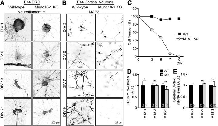
Munc18-1 KO DRG neurons survive in culture without compensatory expression of Munc18-2 or -3. A, DRG cultures form E14 animals of WT and Munc18-1 (M18–1) KO were fixed at different time points (DIV 1, 6, 13, and 21) and stained with neurofilament antibody; no degeneration was observed in Munc18-1 KO DRG neurons. B, Cortical neuronal cultures from E14 animals, WT and Munc18-1 KO, were fixed at different time points and stained for a dendritic marker (MAP2), at DIV 7. Munc18-1 KO neurons were few and underdeveloped. C, Quantification of number of neurons per 97 fields of view showed a decrease in percentage of surviving Munc18-1 KO cells, but not WT cells. D, mRNA levels of Munc18-1, -2, and -3 were quantified in DRG neurons from E14 animals, no changes were observed; Munc18-1 WT: 1.0 ± 0.19 A.U.; KO: 0.0 ± 0.00 A.U.; Munc18-2 (M18-2) WT: 1.0 ± 0.12 A.U.; KO: 1.0 ± 0.06 A.U.; Munc18-3 (M18-3) WT: 1.0 ± 0.07 A.U.; KO: 0.84 ± 0.13 A.U.; not significant, p > 0.05 (Mann–Whitney U test). *p < 0.05 (Mann–Whitney U test). Data are mean ± SEM. E, mRNA levels of Munc18-1, -2, and -3 were quantified in the cerebral cortex from E18 animals; no changes were observed; Munc18-1 WT: 1.0 ± 0.00 A.U.; KO: 0.0 ± 0.00 A.U.; Munc18-2 WT: 1.0 ± 0.00 A.U.; KO 1.1 ± 0.04 A.U.; Munc18-3 WT: 1.0 ± 0.00 A.U.; KO: 1.05 ± 0.21 A.U.; not significant, p > 0.05 (Mann–Whitney U test). *p < 0.05 (Mann–Whitney U test). Data are mean ± SEM.

