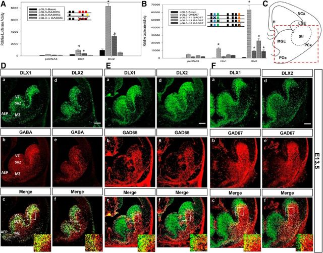
Figure 4.
A, B, Dlx1 and Dlx2 activate transcription of Gad1 (Gad67) and Gad2 (Gad65) reporter constructs in vitro. Transient transfection assays in C6 glioma cells with (A) Gad65 promoter regions i or ii, and (B) Gad67 1.3 kb promoter constructs (contains regions i and ii; Fig. 1A), containing homeodomain binding sites cloned into a pGL3-luciferase reporter construct, were performed in the absence or presence of DLX1 or DLX2 coexpression. DLX1 and DLX2 activate transcription of the reporter genes using the Gad65 and Gad67 promoters, with DLX2 as a more robust activator. Mutations of specific TAAT/ATTA binding motifs within (A) Gad65 or (B) Gad67 promoter sequences lead to a significant reduction of transcriptional activation of these reporter gene constructs. All luciferase activities were relative and normalized to the activity level of the internal control β-gal. Average ± SEM of at least triplicate experiments. *p < 0.05. ΔGAD65i, Mutation of 3 TAATs of Gad65 region I; Δ1GAD67, mutation of the second TAAT of Gad67 region I; Δ2GAD67, mutation of the third TAAT of Gad67 region I; Δ3GAD67, mutation of the fourth TAAT of Gad67 region ii. C–F, Coexpression of DLX homeodomain proteins and the GABA neurotransmitter in wild-type E13.5 basal telencephalon. C, Schematic diagram of coronal section of the E13.5 forebrain, showing basal telencephalon in red dashed box. Sections were double-labeled with specific antibodies against DLX1 (Da, Ea, Fa), DLX2 (Dd, Ed, Fd), GABA or GAD65 or GAD67 (Db, Eb, Fb, De, Ee, Fe) of E13.5 ganglionic eminences. Da, Ea, Fa, Dd, Ed, Fd, DLX1- or DLX2-positive cells (green) in the VZ and SVZ of the LGE, MGE, and AEP. Db, Eb, Fb, De, Ed, Fe, GABA or GAD65 or GAD67-labeled cells (red) in the same tissue sections throughout the basal telencephalon, predominantly in SVZ and MZ of the LGE and AEP. Bottom, The overlay of the two images with GABA/GAD65/GAD67 coexpressed with DLX proteins in most SVZ interneurons (yellow). Scale bar, 200 μm. Insets, Representative of a ∼10× enlargement of the dashed inset box to better demonstrate colabeled cells. H, Hippocampus; LGE, lateral ganglionic eminence; NCx, neocortex; PCx, paleocortex; POa, anterior preoptic area; Str, striatum.

