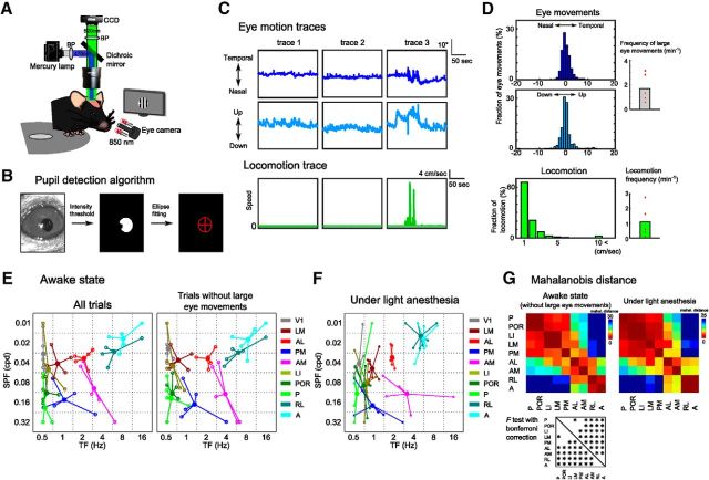
Figure 5.
Spatiotemporal preferences of HVAs were consistent between awake and anesthetized mice. A, Experimental setup. B, Brief explanation of the pupil detection algorithm. We detected the pupil area by intensity threshold and computed the center of the detected pupil area by an ellipse fitting (see Materials and Methods). C, Representative traces of eye movements (top row, horizontal eye movements; middle row, vertical eye movements) and locomotion (bottom row). Although the eye movements were few in traces 1 and 2 (left and middle columns, respectively), large eye movements occurred in trace 3 (right column). We determined trials including large eye movements (>10°) as “trial with large eye movements” (see Materials and Methods). D, Distributions of eye movements (top, horizontal eye movements; middle, vertical eye movements) and locomotion (bottom). Right, Frequencies of large eye movements (>10°) and locomotion. E, Peak points of the fitted response matrix in awake mice (n = 5 mice). The spatiotemporal preferences were similar between the data of all trials and extracted trials without large eye movements. F, Peak points of the fitted response matrix in anesthetized mice. The data are the same as those shown in Figure 3D. G, Colored matrices represent the Mahalanobis distances among HVAs (p < 0.0014, F test with Bonferroni correction). The functional segregation of HVAs in the awake state was very similar to that in the anesthetized state.

