Figure 1.

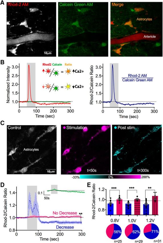
Theta burst neural activity caused a long-lasting reduction in resting free cytosolic Ca2+ in astrocytes. A, Two-photon images of neocortical astrocytes coloaded with the Ca2+ indicator Rhod-2 AM and the morphological dye Calcein Green AM. B, Representative Rhod-2 and Calcein measurements to a single bout of theta burst neural afferent electrical stimulation. Left, Separate Rhod-2 (red) and Calcien (green) traces. Right, Rhod-2/Calcein ratio. C, Images of neocortical astrocytes (left, gray) depicting the transient increase in Ca2+ detected during theta burst activity (middle, magenta), and the drop in Ca2+ in somata and major processes measured following the cessation of theta burst (right, cyan). D, Ca2+ measurements (Rhod-2/Calcein ratio) with evoked theta burst neural activity (0.8 V stimulation) showing two populations of astrocytes: ones that exhibit a significant, transient increase followed by a significant sustained decrease in Ca2+ (blue) and ones that show little response (pink). Inset green trace, Time course summary data of only the Calcein measurement. E, Top, Summary changes in baseline Ca2+ of astrocytes that showed the drop in Ca2+ (blue) and astrocytes that did not show the drop (pink) at different electrical stimulation voltages. Bottom, Pie charts depicting the percentage of cells exhibiting the drop in Ca2+ at different stimulation voltages. **p < 0.01. ***p < 0.001.
