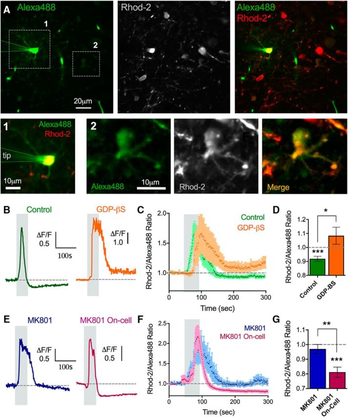
Figure 4.

Glutamate receptors functionally localize to astrocytes. A, Two-photon images showing a whole-cell patched astrocyte and the astrocyte network filled with Alexa-488 (left), costained with Rhod-2 (middle) and the merge (right). Alexa images were gamma filtered at 0.8 to view the less bright coupled astrocytes. 1, Patched astrocyte zoomed in (left image). 2, An astrocyte distant to the patched cell that filled with the Alexa-488 via gap junctional coupling (right 3 images). Ca2+ measurements were made in astrocyte neighbors to the patched cell, such as ROI 2. B, Representative traces of the astrocyte Ca2+ response to theta burst stimulation during a control astrocyte network infusion (green, left) or GDP-β-S loading into the astrocyte network (orange, right). C, Summary traces of the same experiments in B. D, Summary of the peak Ca2+ changes. E, Representative traces of the astrocyte Ca2+ response to theta burst stimulation during the infusion of MK-801 into the astrocyte network (blue, left) or a MK-801 on-cell control (no whole-cell event) (pink, right). F, Summary traces of the same experiments in E. G, Summary of the peak Ca2+ changes. Error bars represent SEM. *p < 0.05. **p < 0.01. ***p < 0.001.
