Figure 4.

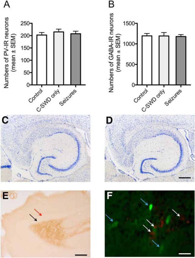
AAV-TeLC injection or related seizures did not induce signs of neurodegeneration. A, B, AAV-TeLC did not affect cell numbers of PV neurons at the injection site. Numbers (mean ± SEM) of PV-positive and GABA-positive neurons were reduced neither in the subiculum of mice that had experienced C-SWD only (n = 6) nor in mice presenting SRSs after AAV-TeLC injection (n = 16). Cell counts were performed 6–8 weeks after AAV-TeLC injection (and monitoring). Numbers are shown for the injected subiculum; they did not differ from those in the contralateral subiculum (data not shown). Controls: uninjected mice (n = 6); data were not different from those for AAV-GFP-injected mice (n = 9; data not shown). C, D, Representative Nissl stains in brain slices at the level of the ventral hippocampus of mice with SRS after AAV-TeLC injection (n = 16; C), and after AAV-GFP injection (seizure free; n = 9; D). E, To test for possible mossy fiber sprouting as a consequence of loss in highly vulnerable mossy cells, we labeled hippocampal mossy fibers by immunohistochemistry for dynorphin. Although mossy fibers were strongly positive for dynorphin, no immunoreactivity was observed in the inner molecular layer of the dentate gyrus, which would be indicative for sprouted mossy fibers (red arrow in E) in AAV-TeLC-injected mice that had experienced SRSs for 6–8 weeks. The black arrow (E) indicates the area of the granule cell layer that is also devoid of dynorphin immunoreactivity (representative for 16 mice). F, Double labeling for the apoptosis marker caspase 3 (red cells, white arrows) and for TeLC/GFP (green cells, blue arrows) at the injection site of AAV-TeLC 10 d after injection. Labeling was performed in three horizontal sections obtained at different dorsoventral levels (∼240 μm apart from each other) from four mice killed 3 or 10 d after AAV-TeLC injection. Zero to maximally five apoptotic cells per section were detected close to the needle track. Note the intact GFP-positive cells expressing TeLC but not caspase 3 in close vicinity to presumably apoptotic cells. As positive controls, we investigated caspase 3 expression in the dorsal hippocampus of mice injected locally with kainic acid 2 and 10 d before (Jagirdar et al., 2015) and observed caspase 3-positive cells at both intervals (data not shown). Scale bars: D (for C, D), 500 μm; E, 200 μm; F, 20 μm.
