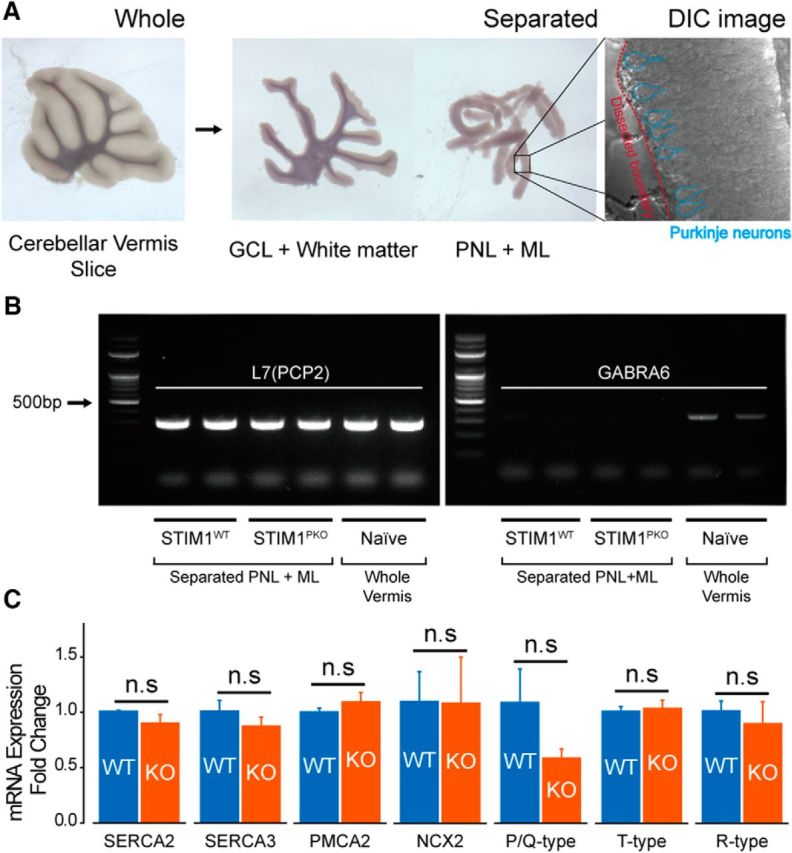
Figure 2.

STIM1 deletion did not significantly change the mRNA expression of Ca2+ clearance/influx sources. A, Slices of the cerebellar vermis (left) were microdissected into PNL + ML and GCL + white matter (middle) according to the visually detectable borderline between PNL and GCL under an illuminated surgery microscope. A DIC microscopic image showed that a piece of separated PNL + ML tissue contained PNs (right). B, RT-PCR (30 cycles) with primers of Purkinje neuronal marker [L7(PCP2)] and granule cell marker (GABRA6) showed that separated PNL + ML showed a lack of genomic contents from granule cells. C, qPCR analysis of representative Ca2+ clearance and influx sources of PNs (number of samples from different animals: wild-type, n = 3; STIM1PKO, n = 3). From left to right: SERCA2 (p = 0.700), SERCA3 (p = 0.700), PMCA2 (p = 0.700), NCX2 (p > 0.999), P/Q-type VGCCs (Cav2.1; p = 0.100), T-type VGCCs (Cav3.1; p > 0.999), R-type VGCCs (Cav2.3; p > 0.999). Mann–Whitney U test was used for C. Error bars denote the SEM.
