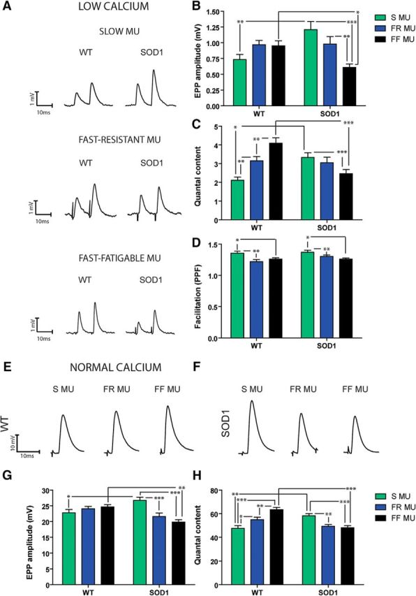
Figure 6.

EPP amplitude and quantal content are increased at P180 in S NMJs but decreased in FF NMJs. A, Typical traces of EPPs evoked by a paired-pulse protocol (0.2 Hz, 10 ms interval) in a low Ca2+/high Mg2+ solution for S (top), FR (middle), and FF NMJs (bottom) of WT and SOD1G37R mice. B–D, Histograms of the mean ± SEM of EPP amplitude (B), quantal content (C), and paired-pulsed facilitation ratio (D) of the S (green), FR (blue), and FF NMJs (black) from WT and SOD1G37R mice. Note that SOD1 NMJs showed a reverse MU pattern. E, F, Example traces of EPPs in physiological Ca2+ concentration with μ-conotoxin GIIIB to block muscle contractions, for WT (E) and SOD1 (F) mice. G, H, Histograms of the mean ± SEM of EPP amplitude (G) and quantal content (H) performed in normal extracellular Ca2+ concentration. Note the similarities between data obtained in low vs normal Ca2+ conditions, confirming the striking synaptic differences observed in the mutant mice. *p < 0.05; **p < 0.01; ***p < 0.001.
