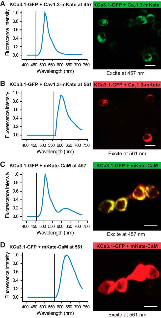
Figure 3.

CaV1.3 does not associate with KCa3.1 at a level that supports FRET. Fluorescence confocal images of tsA-201 cells expressing constructs of KCa3.1-GFP (donor molecule) and CaV1.3-mKate (acceptor molecule) excited with a laser at 457 or 561 nm, respectively. Plots show the excitation line (vertical line) and the associated average emission spectra for each condition obtained from 20–30 cells from three to five independent experiments. A, B, Excitation at 457 nm of cells expressing KCa3.1-GFP and CaV1.3-mKate results in only a GFP emission spectra, indicating no FRET. C, In comparison, the expression of KCa3.1-GFP and mKate-CaM as a positive control revealed FRET, as indicated by an emission spectra for mKate (peaks, ∼630 nm; Fig. 3C) when excited at 457 nm. D, Excitation of cells expressing KCa3.1-GFP and mKate-CaM at 561 nm returns only mKate spectra. Scale bars, 20 μm.
