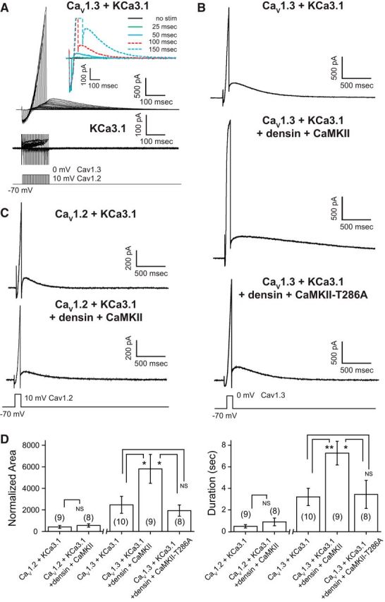
Figure 4.

CaV1.3 channels evoke a long outward tail current when coexpressed with KCa3.1. A, A long outward tail current is preferentially evoked in tsA-201 cells coexpressing CaV1.3 and KCa3.1 channels. Inset of a magnified view of tail currents reveals a graded activation of the tail current in direct relation to the duration of a step command. However, no tail currents are detected in cells expressing KCa3.1 in isolation (bottom records). B, Coexpression of CaV1.3 and KCa3.1 along with densin and CaMKII reveals larger outward tail currents of up to 6 s in response to a 100 ms depolarizing step compared with cells expressing CaV1.3 and KCa3.1 alone. Larger tail currents are not observed in cells expressing the autophosphorylation mutant CaMKII-T286A along with densin, CaV1.3, and KCa3.1. C, Outward tail currents recorded in CaV1.2- and KCa3.1-expressing cells are comparatively small in amplitude and are not augmented by the coexpression of densin and CaMKII. D, Bar plots of the mean area and duration of tail current activated by a 100 ms step. All values are normalized to the preceding inward peak calcium current. CaV1.3 channels are more effective than CaV1.2 in generating an outward tail current that is further augmented by densin and autophosphorylated CaMKII. A one-way ANOVA followed by post hoc unpaired Student's t test was performed between the CaV1.3 groups for area (p < 0.05) and duration (p < 0.05) and an unpaired Student's t test between CaV1.2 groups (NS). NS, Not significant. *p < 0.05; **p < 0.01. Cell numbers are shown in brackets in D.
