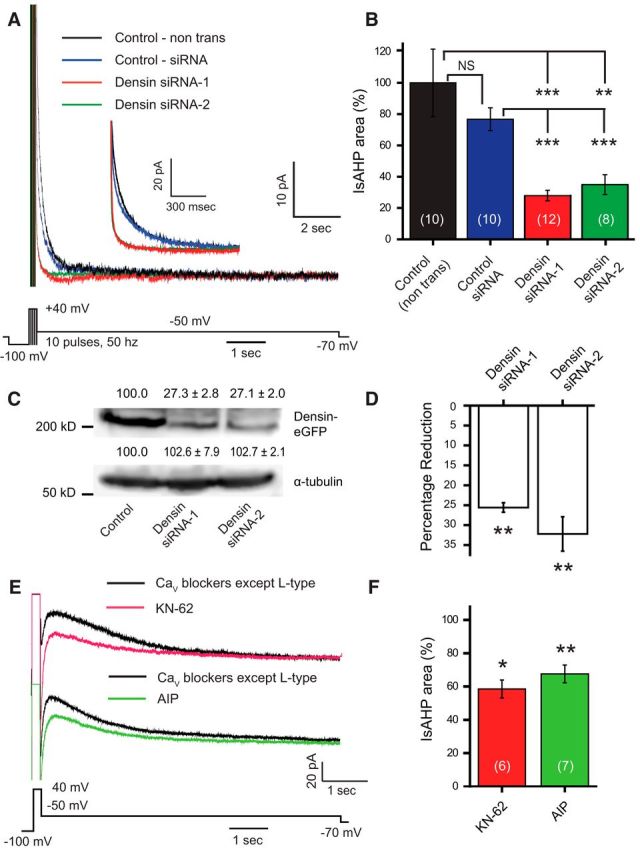
Figure 7.

The IsAHP in hippocampal neurons is regulated by densin and CaMKII. A, Whole-cell recordings of IsAHP evoked by a series of brief pulses to 40 mV in dissociated hippocampal pyramidal neurons in the presence of 100 nm apamin and 10 μm XE-991 to block SK and Kv7 potassium channels, respectively. Pulse trains consisted of 10 5 ms pulses at 50 Hz. Inset shows an enlarged view of the early component of the IsAHP recorded under the indicated conditions. B, The area of the IsAHP is significantly reduced in cells pretreated with either of two siRNAs directed against the postsynaptic scaffolding protein densin compared with nontransfected cells (control) or with cells transfected with a universal control siRNA (one-way ANOVA; p < 0.001). C, Representative Western blot of protein levels of densin or the loading control α-tubulin prepared from lysates of tsA-201 cells expressing densin-GFP with either of two forms of densin siRNA. Proteins were detected using antibodies against GFP or α-tubulin and the density was quantified (using ImageJ) with mean percentages relative to cells expressing only densin-GFP shown above each lane (n = 3; one-way ANOVA, p < 0.001). The levels of densin are greatly reduced in tsA-201 cells cotransfected with either siRNA-1 or siRNA-2 compared with controls. D, Percentage reduction of densin mRNA levels in cultured hippocampal neurons transfected with either of two densin siRNAs compared with cells transfected with control siRNA. The Ct values of densin mRNA from each group are normalized with the Ct value of β-actin as the internal control. Both siRNAs significantly reduced densin mRNA levels in hippocampal neurons compared with neurons transfected with control siRNA (n = 3; one-way ANOVA, p < 0.01). E, Whole-cell recordings of IsAHP in rat CA1 pyramidal cells evoked in a medium containing blockers against all CaV channels except L-type (1 μm ω-conotoxin MVIIC, 200 nm ω-agatoxin IVA, 200 nm SNX-482, and 1 μm TTA-P2). The remaining presumed L-type channel-activated IsAHP is reduced by the CaMKII inhibitors KN-62 and AIP. KN-62 (10 μm) was bath applied, and AIP (20 μm) was internally infused through the electrode. F, Bar plots of mean values for IsAHP area in CA1 pyramidal cells indicate a significant reduction upon the application of CaMKII inhibitors KN-62 or AIP relative to traces obtained before the drug application (as in E; paired Student's t test). *p < 0.05; **p < 0.01; ***p < 0.001. NS, Not significant. Cell numbers are shown in brackets in B and F.
