
Figure 7.
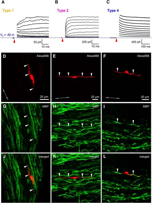
Only SCs without response to glutamate are involved in myelination of axons. A, Example of a current response to a series of depolarizing voltage steps applied from Vhold = −80 mV (blue dotted line) with an increment of +10 mV for a type 1 cell filled with Alexa Fluor-568 (D) and stained for MBP (G, J). The currents are overlaid and the capacitive transients are blanked for clarity. Red arrow indicates the time point when a voltage step started. B, As in A but for a type 2 cell. C, As in A but for a type 4 cell. Note the difference between the scale bars in A–C. D–F, Type 1, type 2, and type 4 cells filled with Alexa Fluor-568 during patch-clamp recordings in the sciatic nerve slice (red, single confocal plane). Arrowheads point to the processes of the cells. G–I, Postrecording immunohistochemistry for MBP (green; Cy5 in G and H or Alexa Fluor-488 in I). The confocal plane and the position of the arrowheads are the same as in D–F. J–L, Overlay of red and green channels shown in D–I. Note that the recorded cells of type 1 and type 2 are negative for MBP, whereas the recorded cell of type 4 is positive for MBP. Gray double-ended arrow indicates the orientation of the axons in the nerve.