
Figure 3.
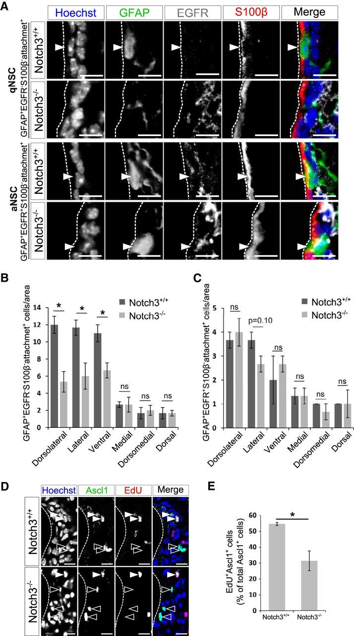
Deletion of Notch3 resulted in domain-specific reduction of qNSCs but not of aNSCs. A, Immunostaining on brain sections of 2-month-old control (Notch3+/+) and Notch3-null (Notch3−/−) mice. GFAP, Green; EGFR, white; S100β, red. qNSCs are defined as GFAP+EGFR−S100β− cells with an apical attachment (attachment+) and aNSCs as GFAP+EGFR+S100β−attachment+ cells. Arrowheads indicate each cell type. B, Quantification of the number of GFAP+EGFR−S100β−attachment+ cells. GFAP+EGFR−S100β−attachment+ cells are decreased in the dorsolateral, lateral, and ventral regions of the SEZ, but not significantly in the medial, dorsomedial, and dorsal regions, which is consistent with other cell types. C, Quantification of the number of GFAP+EGFR+S100β−attachment+ cells. GFAP+EGFR+S100β−attachment+ cells are not significantly reduced in any region of the SEZ. D, Ascl1 (green) and EdU (red). Open and closed arrowheads indicate Ascl1+EdU− cells and Ascl1+EdU+ cells, respectively. E, Percentage of EdU+ cells among Ascl1+ cells. Scale bar, 10 μm. Quantitative data are mean ± SEM. n = 3 and 3. Nuclei were stained with Hoechst (blue). Broken lines indicate the ventricular surface. Two-tailed Student's t test; *p < 0.05, **p < 0.01.