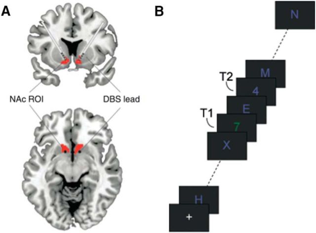
Figure 1.
A, Schematic illustration of deep-brain electrodes in the ventral striatum. Red represents the core of the nucleus accumbens (NAc). Adapted with permission from Figee et al. (2013). B, The AB task. Subjects had to detect two targets (T1 and T2; two numbers) in a rapid stream of distractor stimuli. Shown is an example of a short T1–T2 interval trial.

