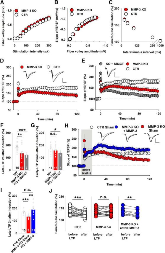
Figure 3.

MMP-3 KO mice had impairments of late LTP in the CA3-CA1 pathway but normal basal excitatory synaptic transmission. A, B, Input–output relationships measured for fiber volley amplitudes (A) (WT, white circles, n = 27 slices; MMP-3 KO, red circles, n = 41 slices) and fEPSPs slopes (B), which were not significantly different between the WT and MMP-3 KO groups (p > 0.2 for all data points, t test). C, Short-term plasticity tested as fEPSP paired-pulse facilitation at various interstimulus intervals, showing no difference between WT and MMP-3 KO mice (WT, n = 20 slices; MMP-3 KO, n = 32 slices; for each interstimulus interval; p > 0.3, t test). D, MMP-3 KO mice had strong deficits in late LTP (CTR: 181 ± 10%; MMP-3 KO: 134 ± 12%; t test, t(30) = 2.94, p = 0.006) but not early LTP (CTR: 170 ± 8%; MMP-3 KO: 157 ± 8%; t test, t(30) = 1.16, p = 0.26) 20 min after induction. Insets, Representative fEPSP traces from WT and MMP-3 KO slices before (gray) and 115–120 min after (black) LTP induction. Stimulation artifacts were removed. Calibration: vertical, 0.5 mV; horizontal, 5 ms. E, SB-3CT application to MMP-3 KO slices further impaired late LTP. MMP-3 KO and WT data were similar to those depicted in Figure 2D. Comparisons of LTP that was induced in MMP-3 KO slices in the presence of SB-3CT with LTP that was induced in WT slices revealed impairments of early LTP (MMP-3 KO+SB-3CT: 138 ± 6%; t test vs CTR, t(20) = 2.67, p = 0.014) and late LTP (MMP-3 KO+SB-3CT: 119 ± 5%; Mann–Whitney U test vs CTR, U(20) = 6.0, p = 0.001). F, G, Summary plots that depict the effects of MMP inhibitors and MMP-3 deficiency on late LTP (F) and early LTP (G). Simultaneous inhibition or KO of both MMP-3 and MMP-9 impaired early LTP, whereas MMP-3 KO affected only late LTP. H, Incubation of MMP-3-deficient slices with exogenous active MMP-3 protein during 100 Hz stimulation (from −5 to 15 min, gray area) rescued the impairment of late LTP (CTR sham-treated: 170 ± 5%; MMP-3 KO sham-treated: 132 ± 6%; MMP-3 KO treated with active MMP-3: 189 ± 13%; t test, CTR vs KO sham, t(17) = 5.1, p < 0.001; t test, KO sham vs KO treated, t(18) = 5.1, p = 0.002; Mann–Whitney U test, CTR vs KO treated, U(19) = 33, p = 0.13). I, Summary plot that depicts the effects of infusing the exogenous active MMP-3 protein on late LTP recorded in MMP-3 KO slices. The short exposure to MMP-3 activity restored the magnitude of late LTP in MMP-3-deficient slices. J, Comparison of short-term plasticity before and 2 h after LTP induction. In WT slices, the induction of LTP significantly decreased the paired-pulse facilitation (n = 21 slices, paired t test, p < 0.001). The impairment of late LTP in MMP-3 KO slices was accompanied by a lack of change in the paired-pulse facilitation after LTP (n = 20 slices, paired t test, p = 0.68). Brief infusion of active MMP-3 protein during LTP induction in MMP-3-deficient slices restored the changes in short-term plasticity accompanying LTP that were observed in WT slices (n = 9 slices, paired t test, p = 0.006). *p < 0.05. **p < 0.01. ***p ≤ 0.001. n.s., not significant.
