Figure 1.
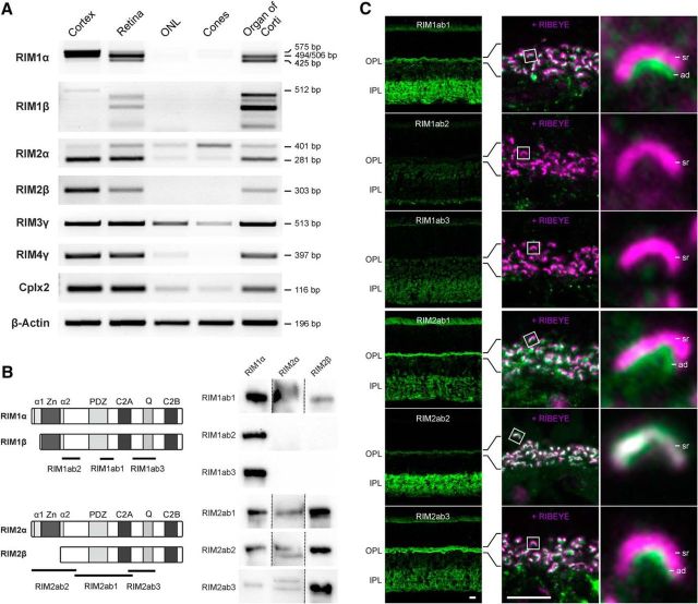
Expression and distribution of RIM family members at photoreceptor ribbon synapses. A, RT-PCR of cDNA from cortex, whole retina, ONL, isolated cone photoreceptors (Cones), and organ of Corti with specific primers for the different RIM isoforms. As a control for the purity of the photoreceptor samples, primers for Complexin2 (Cplx2) were used and RT-PCR with β-Actin primers served as loading control. B, Left, Schematic representation of RIM1α/β and RIM2α/β and epitope locations of the used antibodies. Right, Western blots of overexpressed RIM1α, RIM2α, and RIM2β stained with the different RIM antibodies to test for antibody specificity. C, Left, Images of vertical sections through the C57BL/6 mouse retina stained with the different RIM antibodies. Right, Immunocytochemical double staining of the OPL with the different RIM antibodies (green) and with an antibody against RIBEYE (magenta). α1/α2, RAB3A-binding domains; ad, arciform density; bp, base pairs; C2, C2 domain; PDZ, PDZ domain; Q, glutamine-rich heptad repeat; sr, synaptic ribbon; Zn, zinc finger. Scale bar in C, 5 μm.

