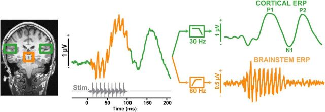
Figure 2.
Dual brainstem and cortical neuroelectric recording paradigm. Schematic derivation of brainstem FFR (orange) and cortical ERP (green) responses from grand averaged speech-evoked activity via high- and low-pass filtering, respectively. Note that time is not to scale in the right traces. Gray trace, stimulus waveform. MRI anatomy illustrates the presumed source generators of brainstem and cortical potentials. In the current experiment, brainstem and cortical responses were recorded serially and isolated via this selective filtering technique.

