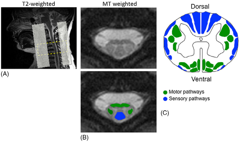
Figure 1.
(A) T2-weighted sagittal localizer demonstrating typical patient positioning and the slice prescription for MT weighted acquisition, covering C4–C6 vertebral levels. Saturation bands were set ventrally and dorsally to limit aliasing and ghosting artifacts. (B) Anatomically defined ROIs on the MT-weighted image (for MTR) were selected over the ventromedial and dorsolateral (green) descending motor pathways and the dorsal columns (blue) of the cervical spinal cord. (C) Motor and sensory pathways.

