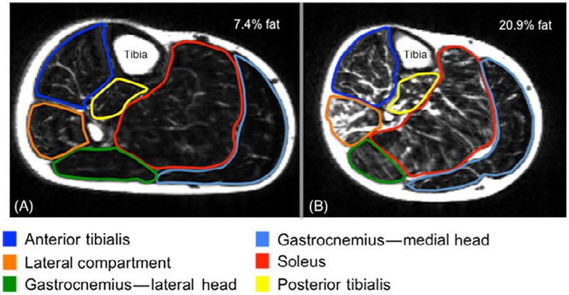
Figure 2.
MRI (fat-only image) of the right plantar/dorsiflexors in (A) recovered subject and (B) severe whiplash subject. Note the increased signal throughout the plantar/dorsiflexors in the severe whiplash subject(B) suggestive of fatty infiltrates. This is not observed in the recovered whiplash subject (A).
NB. The data for the recovered subject (A) (at 3 months post-injury event) was presented solely for visual observation of and comparison with the lower extremity muscles in this chronic subject (B).

