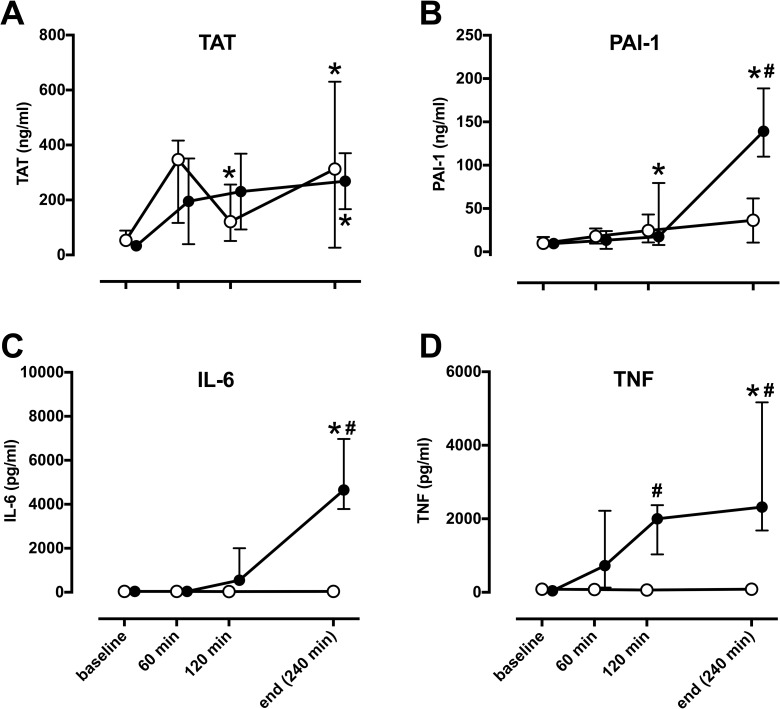
Fig 2. Sepsis but not pulmonary artery banding induces pro-inflammatory changes in plasma.
In comparison to baseline, E. coli induced sepsis (filled circles) significantly increased markers of coagulation TAT (A) and PAI-1 (B) as well as cytokines IL-6 (C) and TNF (D), while pulmonary artery banding (open circles) lead to significant increase of TAT only (*). Significant differences between sepsis and pulmonary artery banding (#) were visible in all markers except for TAT. TAT; thrombin-antithrombin complex, PAI-1; plasminogen activator inhibitor-1, IL-6; interleukin-6, TNF; tumor necrosis factor. All values Median ± Interquartile Range. Generalized linear mixed model with post-hoc comparison to baseline (*) and pairwise comparison between pulmonary artery banding and sepsis at each time-point (#). Bonferroni correction for multiple testing; p< 0.05.

