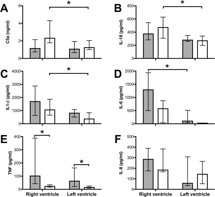
Fig 3. Sepsis and pulmonary artery banding induce inflammation in RV myocardium.
Tissue samples from RV and LV at the end of the experiment were analyzed for C5a (A), IL-18 (B), IL-1β (C), TNF (D), IL-6 (E), and IL-8 (F). (A-C) C5a, IL-18 and IL-1β were significantly induced during pulmonary artery banding (open bars) in the RV compared to the LV. (D) IL-6 was significantly increased during sepsis (filled bars) in the RV compared to the LV. (E) TNF was significantly increased in both ventricles during sepsis only and not by pulmonary artery banding, while (F) IL-8 was not significantly different between ventricles nor between treatments. All values Median ± Interquartile Range. Mann-Whitney U test, *; p < 0.05.

