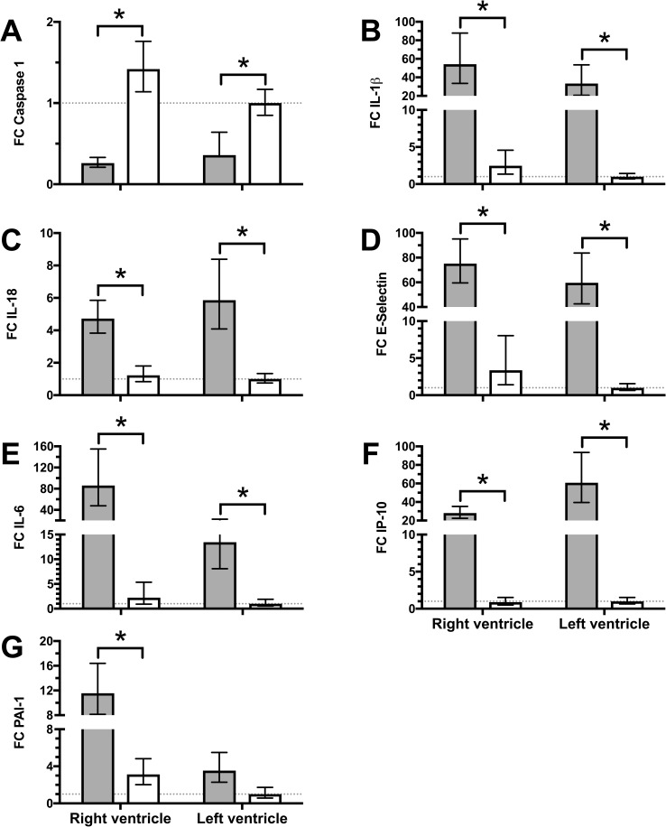
Fig 4. Myocardial RNA-expression differs between sepsis and pulmonary artery banding.
Tissue samples from the LV and RV were obtained at the end of the experiment and analyzed for RNA-expression. LV of animals with pulmonary artery banding (open bars) served as control (indicated by dotted line). Sepsis (filled bars) led to a significant decrease of caspase-1 expression (A), while IL-1β increased significantly (B). IL-18, E-Selectin, IL-6, and IP-10 (C-F) increased significantly more during sepsis compared to pulmonary occlusion in both RV and LV. PAI-1 (G) did only increase significantly in RV during sepsis compared to pulmonary occlusion. No statistically differences were found between ventricles during sepsis or pulmonary occlusion. FC; Fold Change, all data presented as Mean ± 95% confidence interval, 1-way ANOVA with post-hoc all pairwise comparison and Bonferroni correction, *; p < 0.05.

