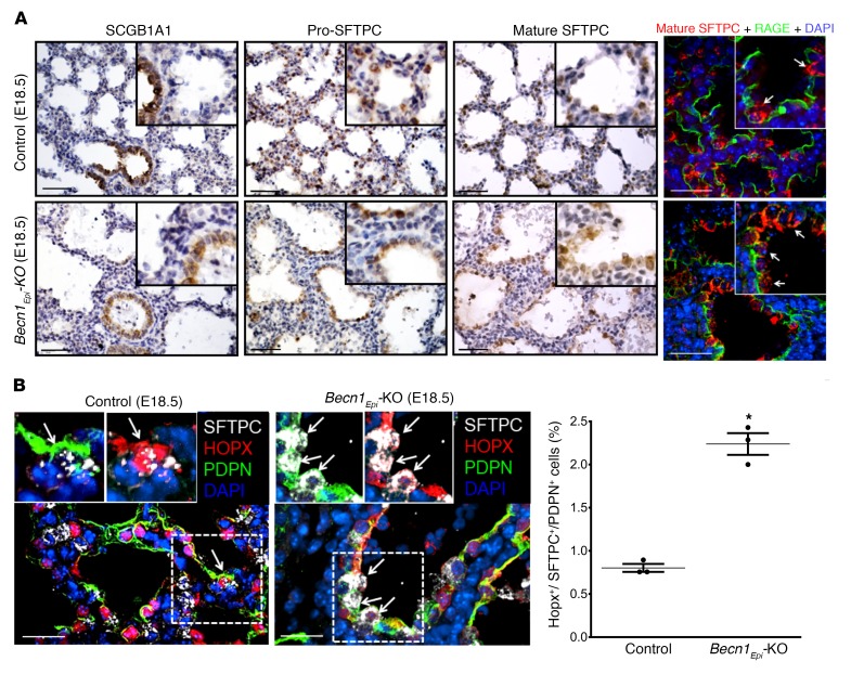
Figure 10. Conditional deletion of Becn1 delays distal epithelial differentiation.
(A) Representative IHC images for Clara cell secretory protein (SCGB1A1), pro-SFTPCC, and mature SFTPC expression in lung tissue sections from E18.5 Becn1Epi-KO and littermate control fetuses. Scale bars: 50 μm; original magnification, ×20 (insets). IF microscopic images show lung tissue sections from E18.5 Becn1Epi-KO and littermate control fetuses stained for mature SFTPC (red) and RAGE (green). The white arrows in the insets point to cuboidal alveolar type II epithelial cells. Scale bars: 25 μm; original magnification, ×40 (insets). (B) Confocal IF microscopic images of E18.5 lung tissue from Becn1Epi-KO mice costained for SFTPC (white), HOPX (red), and PDPN (green). Nuclei were stained with DAPI. Arrows indicate alveolar precursor cells detected by an overlap of all these markers. Scale bars: 25 μm; original magnification, ×40 (insets). Graph indicates the percentage of alveolar precursor cells that stained positive for SFTPC, HOPX, and PDPN in E18.5 lung sections from Becn1Epi-KO and littermate control mice. Data are expressed as the mean ± SEM (n = 3 separate experiments). *P < 0.05 versus WT control, by Student’s t test.

