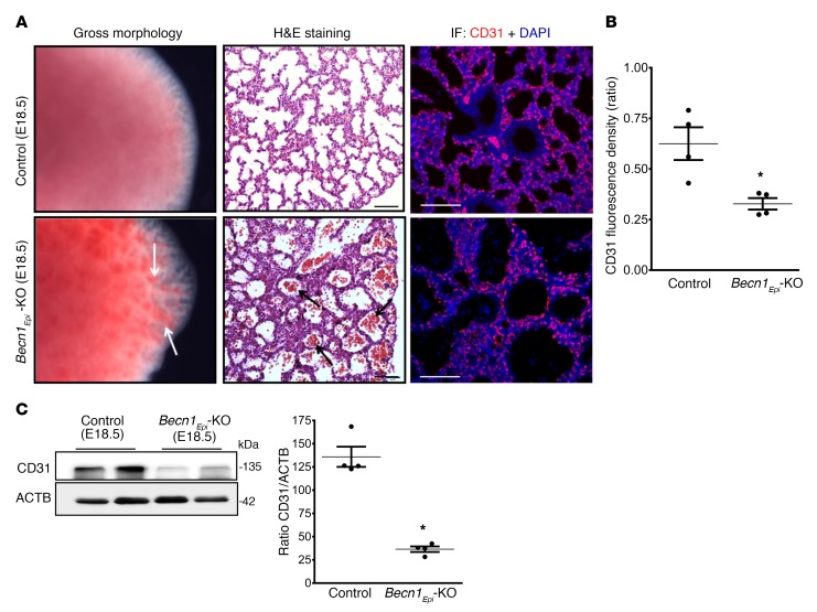
Figure 8. Conditional deletion of Becn1 alters proper pulmonary vascular development.
(A) Left panel: Gross morphology of E18.5 lungs from control (WT) and Becn1Epi-KO embryos. White arrows point to hemorrhage regions in Becn1Epi-KO lung. Middle panel: Representative light photomicrographs of H&E-stained lung sections from E18.5 littermate control (WT) and Becn1Epi-KO mice. Note the infiltration of red blood cells in the enlarged air spaces (black arrows) in Becn1Epi-KO lung. Right panel: Confocal IF microscopic images of embryonic lungs (E18.5) stained for the endothelial cell marker CD31 (red). Nuclei were stained with DAPI (blue). Scale bars: 100 μm (middle panel) and 50 μm (right panel). (B) Quantification of the CD31/DAPI fluorescence ratio of E18.5 lungs from control (WT) and Becn1Epi-KO embryos. Data represent the mean ± SEM (n = 3 separate lungs). *P < 0.05 versus WT control, by Student’s t test. (C) Representative immune blot for CD31 on whole E18.5 lung lysates from control (WT) and Becn1Epi-KO embryos. The membrane was re-used in Figure 7C and Figure 8C, which show the same loading control. Graph shows densitometric analysis of CD31 expression. ACTB was used as a protein loading control. Data represent the mean ± SEM (n = 4 separate lungs). *P < 0.05 versus WT control, by Student’s t test.

