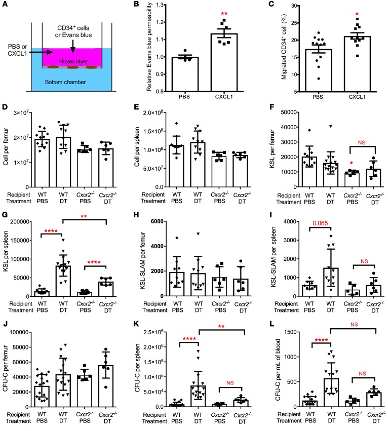
Figure 6. CXCR2 signaling contributes to HSPC mobilization following bone marrow DC ablation.
(A–C) Human umbilical vein endothelial cells (HUVECs) cultured in a transwell to establish a monolayer were treated with PBS or CXCL1. (B) Relative amount of Evans blue dye in the lower chamber after 1 hour of CXCL1; data are normalized to PBS-treated samples (n = 5 or 6 mice). (C) Human bone marrow CD34+ cells were seeded on the HUVEC monolayer and treated with CXCL1 for 24 hours; the lower chamber contained 100 ng/ml CXCL12. Shown is the percentage of CD34+ cells that migrated to the lower chamber (n = 11–12 replicates). (D–L) Bone marrow cells from Zbtb46dtr mice were transplanted into irradiated WT or Cxcr2–/– recipients. Eight weeks after transplantation, mice were treated with PBS or DT for 6 days. Total cellularity of bone marrow (D) and spleen (E), the number of KSL cells in bone marrow (F) and spleen (G), the number of KSL-SLAM cells in bone marrow (H) and spleen (I), and the number of CFU-Cs in bone marrow (J), spleen (K), and blood (L) is shown (n = 6 per cohort). The WT recipient data are the same as shown in Figure 2. Data are mean ± SEM. *P < 0.05; **P < 0.01; ****P < 0.0001 compared with the PBS-treated group, unless otherwise specified. Significance was determined using an unpaired Student’s t test for B and C, or ANOVA with Tukey’s Honest Significant Difference post hoc analysis for D–L.

