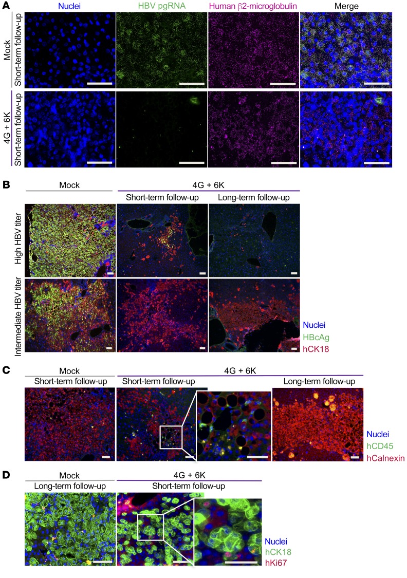
Figure 6. In situ analysis of antiviral effects of TCR-grafted T cells in humanized livers.
Liver tissue from HBV-infected mice treated with mock-transduced T cells or 2 × 10⁶ HBV-specific T cells (1 × 10⁶ with 6KC18 plus 1 × 10⁶ with 4GS20) were used from days 18–19 (short-term follow-up) or day 55 after T cell transfer (long-term follow-up). (A) RNA in situ hybridization for HBV pgRNA (green) and human β2-microglobulin (magenta) against nuclei staining (blue) was performed to determine the occurrence of HBV-specific RNA transcripts. (B) As indicated, mice were categorized as having a high (109 to 1010 copies/ml) or low (107 to 108 copies/ml) HBV titer. Representative immunohistochemical staining for cell nuclei (blue), HBV core protein (green), and human CK18 (hCK18) (red). (C) Human CD45+ cells (green) were stained against human calnexin (hCalnexin) (red) as a marker for human hepatocytes and cell nuclei (blue). (D) To determine potential proliferation in mice treated with T cells, staining for cell nuclei (blue), human CK18 (green), and human Ki67 (hKi67) (red) was done. Scale bars: 50 μm.

