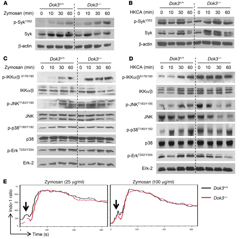
Figure 4. Dok3 deficiency enhances NF-κB and JNK signaling.
(A and B) Immunoblot analysis of p-SykY352 and Syk in purified Dok3+/+ and Dok3–/– neutrophils stimulated for various times with (A) zymosan (10 μg/ml) or (B) HKCA (MOI 1:1). β-Actin served as loading control. Images are representative of 3 independent experiments. (C and D) Immunoblot analysis of p-IKKα/βS176/180, IKKα/β, p-JNKT183/Y185, JNK, p-p38T180/Y182, p38, p-ErkT202/Y204, and Erk-2 in purified Dok3+/+ and Dok3–/– neutrophils stimulated with (C) zymosan (10 μg/ml) or (D) HKCA (MOI 1:1) for indicated periods of time. Images are representative of 3 to 4 independent experiments. Quantifications of immunoblots are shown in Supplemental Figure 4. (E) Calcium signaling in Indo-1–loaded Dok3+/+ and Dok3–/– neutrophils after stimulation with either 25 μg/ml or 100 μg/ml of zymosan. Intracellular calcium flux was monitored in real time via flow cytometry. One representative out of 3 independent experiments is shown.

