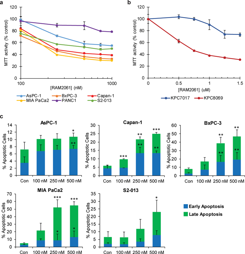
Fig. 3. Treatment of PDAC cells with GGDPSi triggers apoptotic cell death.
a and b MTT assays were performed following a 72-hour incubation period with RAM2061 in six human (a) and two mouse (b) PDAC cell lines (n=4, data are displayed as mean ± stdev). c Human PDAC cells were treated with or without RAM2061 for 72 hours. Cells were stained with fluorescently conjugated Annexin V and propidium iodide (PI) and analyzed by flow cytometry. Data are expressed as the average percentage of Annexin V+/PI- (early apoptotic) and AnnexinV+PI+ (late apoptotic) (n = 3, data are displayed as mean ± stdev, *denotes p < 0.05. **denotes p < 0.01. ***denotes p < 0.001).

