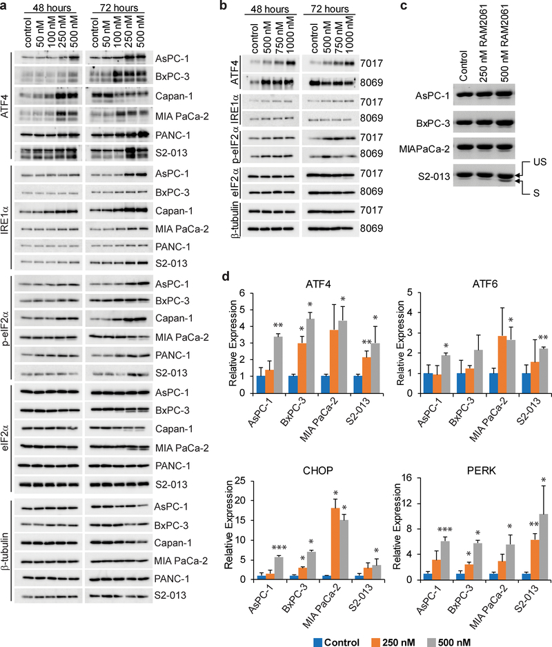
Fig. 4. GGDPS inhibition activates the UPR pathway.
a and b Immunoblot analysis showing protein levels of UPR markers in six human (a) and two mouse (b) PDAC cell lines treated with or without RAM2061 for 48 or 72 hours. Mouse KPC8069 cells are denoted as 8069 and KPC7017 are denoted 7017. β-tubulin is shown as a loading control. Immunoblots are representative of three independent experiments. c Human PDAC cells were incubated for 48 hours with or without RAM2061. PCR was performed using XBP-1-specific primers. The upper band represents unspliced XBP-1 (US) and the lower band represents spliced XBP-1 (S). d qRT-PCR analysis of ATF4, ATF6, CHOP and PERK expression in human PDAC cells incubated in the presence or absence of RAM2061 (48 hour incubation). Data represents fold change normalized to control (n = 3, data are displayed as mean ± SEM, *denotes p < 0.05. **denotes p < 0.01. ***denotes p < 0.001).

