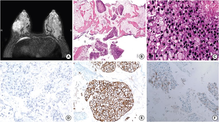
Figure 1. Magnetic resonance imaging and microscopic findings before neoadjuvant treatment. (A) Axial T2-weighted image shows a well-enhanced mass in the right breast. (B) Core biopsy specimen shows tumor cell clusters floating in extracellular mucin pools (hematoxylin and eosin, magnification × 40) (C) Tumor showed extensive signet ring cell differentiation with high grade nuclear features, frequent mitosis, and tumor necrosis (hematoxylin and eosin, magnification × 400) (D, E, F). Tumor cells were negative for ER (D), positive for HER2 (E), and focally expressed GCDFP-15 (magnification × 200).
ER = estrogen receptor; HER2 = human epidermal growth factor receptor 2; GCDFP-15 = gross cystic disease fluid protein 15.

