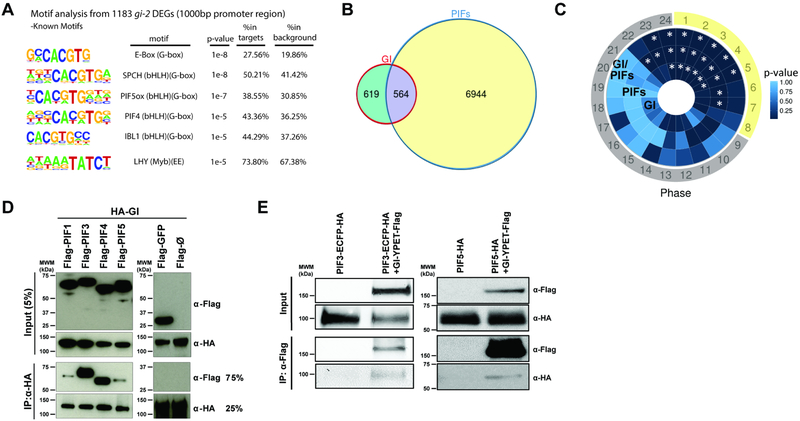
Figure 1. GI shares targets with light signaling components and interacts with PIF proteins.
(A) Over-represented cis motifs at the promoter regions of gi-2 DEGs.
(B) Overlap between DEGs in gi-2 and a comprehensive set of PIF-regulated genes (intersection p<2.2e-16).
(C) Phase enrichment heatmap depicting the p-value of the phase of peak expression enrichment (count/expected) of genes differentially expressed in gi-2 only (GI), regulated by PIFs only (PIFs), and potentially regulated by GI and PIFs (GI-PIFs) under SD conditions (*p<0.01). Day period is marked in yellow and night period in gray.
(D) In vitro pull-down assays showing the interaction between GI and PIFs (PIF1, PIF3, PIF4, and PIF5).
(E) In vivo co-immunoprecipitations in Arabidopsis transgenic seedlings expressing GI-YPET-Flag and PIF3-ECFP-HA (left panel), and GI-YPET-Flag and PIF5-HA (right panel) tagged protein versions.

